

Anatomy of haemorrhoids
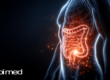
Haemorrhoids are small cushions of spongy tissue located in the anal canal, composed of a rich network of capillaries, veins and arterioles, supported by connective tissue and smooth muscle fibres. They play a fundamental role in the continence mechanism, as they work in conjunction with the anal sphincters and contribute to the control of stool and gas. Moreover, during defaecation, they become engorged and then decongested, modulating their volume through the inflow and outflow of blood. This process reduces the mechanical trauma associated with the passage of stool and protects the structures of the anal canal. From an anatomical perspective, they are divided into:
- Internal haemorrhoids: located within the anal canal, above the so-called dentate (or pectinate) line. They generally do not cause pain, as the mucosa in this area has limited sensitivity. The most common symptom is bleeding.
- External haemorrhoids: located below the dentate line, at the level of the external anal orifice. As they are covered by richly innervated skin, they are sensitive and may cause pain, particularly in cases of inflammation or thrombosis.
Haemorrhoidal disease
When there is an alteration of the normal supporting system of the haemorrhoidal cushions and an increase in pressure within the haemorrhoidal plexus, vascular congestion occurs, leading to inflammation, itching, prolapse, bleeding or the formation of thrombi (blood clots within the haemorrhoids). In such cases, this condition is referred to as haemorrhoidal disease. It is a multifactorial condition, resulting from a combination of predisposing and triggering factors, including chronic constipation or diarrhoea, repeated straining during defaecation, prolonged sitting, pregnancy, obesity, and unhealthy dietary and lifestyle habits—particularly a diet low in fibre and fluids, smoking, and alcohol consumption.
Classification of haemorrhoidal disease
In the presence of symptoms suggestive of haemorrhoidal disease, it is essential to undergo a proctological examination. The specialist, through medical history and physical examination (possibly supplemented by anoscopy), will establish the correct diagnosis and determine the severity of the disease. The most commonly used system for identifying haemorrhoidal disease is the Goligher classification, which categorises internal haemorrhoids into grades:
- Grade I: haemorrhoids may bleed but do not prolapse externally. They are visible only on proctoscopy.
- Grade II: haemorrhoids prolapse during defaecation but return spontaneously.
- Grade III: haemorrhoids prolapse during defaecation or minimal straining and must be manually reduced by the patient.
- Grade IV: permanent, non-reducible prolapse. Oedema, thrombosis, ulceration or incarceration may be present.
Treatment
The classification of haemorrhoidal disease is essential for determining the most appropriate treatment. The available approaches are divided into:
- Conservative treatment: primarily indicated for patients with Grade I and II haemorrhoidal disease and mild to moderate symptoms. It is mainly based on lifestyle and dietary modifications, aimed at reducing haemorrhoidal congestion and preventing straining during defaecation. This includes a high-fibre diet, adequate fluid intake, limiting irritant foods and alcohol, proper intimate hygiene using specific cleansers, and, when necessary, topical or systemic pharmacological therapy.
- Minimally invasive (outpatient) treatment: mainly indicated in Grade I and II cases and, in selected instances, Grade III. It includes instrumental techniques performed on an outpatient basis with the aid of an anoscope, without the need for hospitalisation. The aim is to reduce blood flow to the haemorrhoidal cushions, leading to their gradual shrinkage. The main methods include rubber band ligation, sclerotherapy, photocoagulation and cryotherapy.
- Surgical treatment: generally reserved for Grade III and IV haemorrhoidal disease, or for Grade II cases with significant symptoms that are unresponsive to conservative and outpatient treatments. Surgical techniques may vary depending on the clinical picture and patient characteristics, as well as the type of anaesthesia used, and aim at the removal or definitive reduction of haemorrhoidal prolapse.
In the next article, we will focus in particular on two widely used and effective outpatient treatments: rubber band ligation of haemorrhoids and the sclerotherapy technique, with special emphasis on the combined sclerobanding approach. We will analyse in detail their indications, procedures, benefits and recovery times.
WEBSITE REFERENCES
- Sapi Med dedicated to patients
- SICCR – Italian Society of Colorectal Surgery (ETS)
- Mondadori Digital S.p.A.